The hip joint is a ball and socket type of synovial joint that connects the pelvic girdle to the lower limb. In this joint, the head of the femur articulates with the acetabulum of the pelvic (hip) bone. The joint is very stable and transmits the entire weight of upper body to legs.It also allows a wide range of motion being a multiaxial joint. The articular cartilage covering the ball and socket works as a cushion between the two surfaces.
Normal and arthritic hip
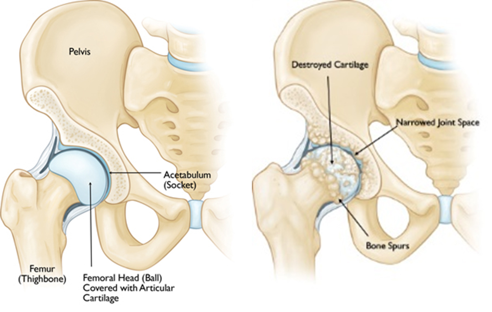
In patients with degenerative arthritis or other hip disorders this cartilage acting like cushion gets destroyed and causes friction in movement of hip joint causing pain in walking and other load bearing activites. The joint may also be damaged in fractures, dislocations ,AVN and inflammatory arthritis. The diseased hip is then replaced with metal and plastic components serving as artificial cushions and allowing painfree movement of hip joint.
Acetabular cup and femoral stem used in THR

Types of Hip Replacement :
The hip replacement material may be cemented using a special kind of cement to hold the cup and stem in place or uncemented using textured or special coating to anchor the implant to the bone. The femoral head may be of cobalt chrome, ceramic or oxynium coated to decrease wear of the polyethylene liner.
Hip replacement is a very rewarding surgery and given the patient a painfree mobile joint again. However realistic expectations are necessary. Patient can recover to the point where he was before the disease process began. You can even jog or walk fast with the artificial joint but should be in a good physical shape to do that. High impact activities are discouraged as they cause accelerated wear and loosening of the prosthetic like any other machinery. Also the patient should not sit or squat on ground as it may cause dislocation of the joint.
SURGERY:
Surgery is generally done in spinal anaethesia and takes a few hours to perform. The surgery is performed using reduced tissue trauma technique using a minimally invasive technique that decreases postoperative pain and causes early recovery for the patient.
Like any surgery it may have complications related to anaesthesia, bleeding , infection, blood clots in the legs or lungs, dislocation, revision surgery, nerve injury causing numbness or weakness. However with proper precautions the complication rate is below 5%.
After the surgery patient may be discharged after 4-5 days after surgery. Depending on the type and patient bone strength he or she may be allowed partial weight bearing at this time or later. The stitches are generally removed at 15 days. Physiotherapy is started on Day 2 after surgery and gradually progressed to walking with walker and with cane by the end of 1 month depending on patient age and strength. Physiotherapy is continued till the patient gains full movement and strength of the leg.